SAS Early Career Interest Group
About Us:
In 2021, the Society for Applied Spectroscopy (SAS) created an Early Career Interest Group (ECIG) and membership category to serve scientists who have graduated with their final degree (bachelors, masters, or doctorate) within the last ten years. The SAS-ECIG is a diverse committee that is dedicated to providing members with a unique experience to help promote and succeed in their career. Together, we aim to support the professional development of early career scientists through award schemes as well as opportunities in leadership, outreach, networking, mentorship, volunteering, formal certification, and employment.
Mission Statement:
The Society of Applied Spectroscopy Early Career Interest Group (SAS-ECIG) aims to support the professional development of early career scientists through award schemes, as well as opportunities in leadership, outreach, networking, mentorship, volunteering, formal certification, and employment.
The primary objectives of SAS-ECIG are to:
- Offer funding opportunities to build, support and promote early career SAS members
- Maintain a socially supportive early career community
- Support early career professional development through mentorship, research collaborations, career development support, and publication opportunities
- Recognize and promote the professional accomplishments and community contributions of early career professionals
Committee Members
|
Fay Nicolson, Founding Chair
|
Work Title: Assistant Professor
Number of Years as a SAS Member: 9
Biggest benefit of SAS: Networking and social events
What did you want to be when you were a child? Musical theater performer
What made you fall in love with spectroscopy? Seeing it be used in real world situations
What is your favorite part of your career or job? Being in an Institute with diverse disciplines and a huge focus on translational research that directly improves the lives of patients
Which scientist, past or present, would you like to meet and why? Marie Skłodowska-Curie. She was relentless in her passion for science during a time when women were rarely allowed in laboratories, let alone recognized for their contributions. Her groundbreaking discoveries in radioactivity opened entirely new avenues in physics and chemistry and laid the foundation for critical advancements in modern medicine today.
|
|
Sam Mabbott, Committee Member
|
Work Title: Assistant Professor
Number of Years as a SAS Member: 12
Biggest benefit of SAS: Networking events at conferences, especially SciX.
What did you want to be when you were a child? Pirate
What made you fall in love with spectroscopy? The breadth and depth of utility that spectroscopies offer.
What is your favorite part of your career or job? Mentoring and collaborating with postdocs, graduate, and undergraduate students. It’s incredibly rewarding to support their growth and share in the discovery process together.
Which scientist, past or present, would you like to meet and why? Leo Szilard, stands out not just for his monumental contributions to physics, but also for his broader moral and philosophical reflections, making him an intriguing figure to learn from on multiple levels.
|
Heather Juzwa, Committee Member |
Work Title: Senior Field Sales Engineer
Number of Years as a SAS Member: 15
Biggest benefit of SAS: I LOVE the SAS people - welcoming, encouraging, and fun!
What did you want to be when you were a child? I was convinced I’d be a teacher. I used to make lesson plans for the summer and make my little sister go to “basement school” every day. Poor girl!
What made you fall in love with spectroscopy? Looking back, I had the best mentors for spectroscopy. Bill Snyder for UV, Ruth Sonoff for Fluorescence, and Ellen Miseo for FTIR. These folks not only had a profound impact on my life but they also gave me a passion for spectroscopy that continues to this day.
How did you get your current position? As a Senior at Pitt, I was a student aide working in the Employment Bureau at Pittcon. At that time, everything was paper, and all the HR representatives were telling me to be in sales as I shuffled resumes and scheduling cards around the bureau. I was adamantly against this because sales reps annoy me. But here I am, almost 25 years later, still a sales rep. The difference with me is that I don’t feel like a sales rep at all. I feel like a problem solver, a customer advocate, and a vessel to promote science anywhere I can!
Which scientist, past or present, would you like to meet and why? Marie Curie has always fascinated me. I admire Curie not just for her groundbreaking scientific discoveries, but for her independence and relentless dedication to her research
|
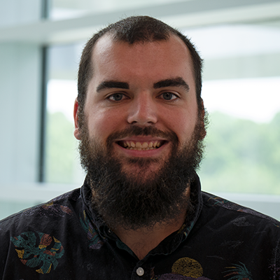
Anthony Stender,
Committee Member |
Work Title: Research Core Manager (Microscopy)
Number of Years as a SAS Member: 10
Biggest benefit of SAS: Mentoring Early Career members
What did you want to be when you were a child? Either a back-up singer for Elvis Presley or an astronaut
What made you fall in love with spectroscopy? The satisfaction I felt during grad school, working on a microscope until 2 AM, switching filters by hand and collecting images of plasmonic nanoparticles, night after night, followed by discovering the patterns that emerged from the data.
What is your favorite part of your career or job? I love working with imaging data and seeing the numbers hidden within the images. And I love those now rare opportunities of working on a microscope, late into the night, especially when I get to share those moments with colleagues.
Which scientist, past or present, would you like to meet and why? Blaise Pascal. Like me, he was part mathematician, a theorist and experimentalist, and a meteorologist. And maybe he could teach me a few things about probability.
|
|
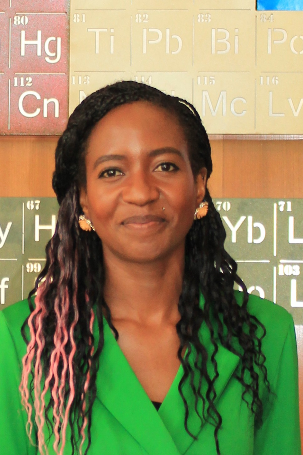
Beauty Chabuka, Committee Member
|
Work Title: PhD. Student
Number of Years as a SAS Member: 5
Biggest benefit of SAS: After completing my master's program, I joined SAS-EC to connect with a global community of spectroscopy professionals. My involvement on the SAS-EC executive board and participation in SAS-EC online events inspired my decision to pursue a PhD. SAS-EC events have provided roadmaps for professional development, highlighting areas for opportunities in both academia and industry.
What did you want to be when you were a child? Doctor
What made you fall in love with spectroscopy? Started in organic chemistry. This passion deepened during my microplastics research, where I combined machine learning with IR spectroscopy to uncover hidden patterns in complex data.
What is your favorite part of your career or job? As a computational chemist, there's something deeply satisfying about watching theoretical peaks align with experimental data. Being able to bypass experimental challenges and using computations to predict molecular properties, reaction mechanisms, and energetics.
Which scientist, past or present, would you like to meet and why? It would be Howard Elliot Zimmerman. His pioneering work in photochemistry laid the foundation for my current research in pericyclic reactions and mechanistic photochemistry. As a second-degree connection in our chemistry family tree, I feel a profound connection to his scientific legacy.
|
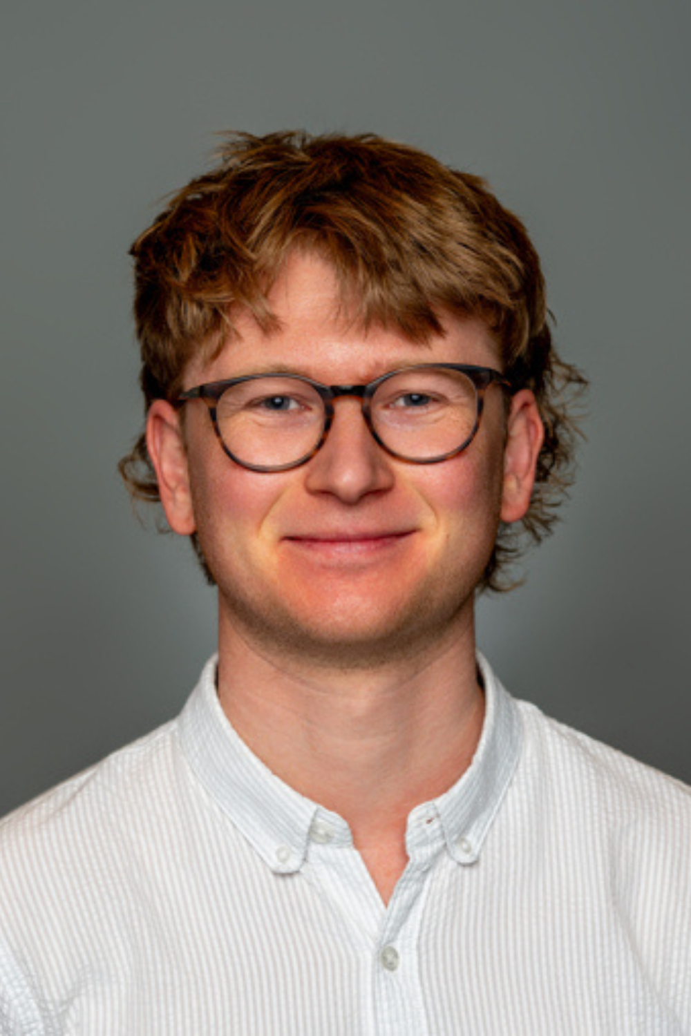
William Skinner, Committee Member |
Work Title: Postdoctoral research fellow
Number of Years as a SAS Member: 2
Biggest benefit of SAS: The supportive and fun applied spectroscopy community and the networking opportunities that come with it.
What did you want to be when you were a child? Inventor
What made you fall in love with spectroscopy? The creative and interdisciplinary research opportunities in applied spectroscopy. I've particularly enjoyed working with chemists, physicists, and clinicians to answer biological questions.
What is your favorite part of your career or job? The moment when something finally clicks and I understand the system that I'm working with at a deeper level. Whether it is troubleshooting or a broader, more universal understanding of a physical or biological process.
Which scientist, past or present, would you like to meet and why? Leonhard Euler. He laid the foundation for so much of what we take for granted and rely on in science, from the way we express mathematical equations to the design of microscopes. I'd like to have a chat with him and get an idea of what one of the foundational minds of modern science was like.
|
|
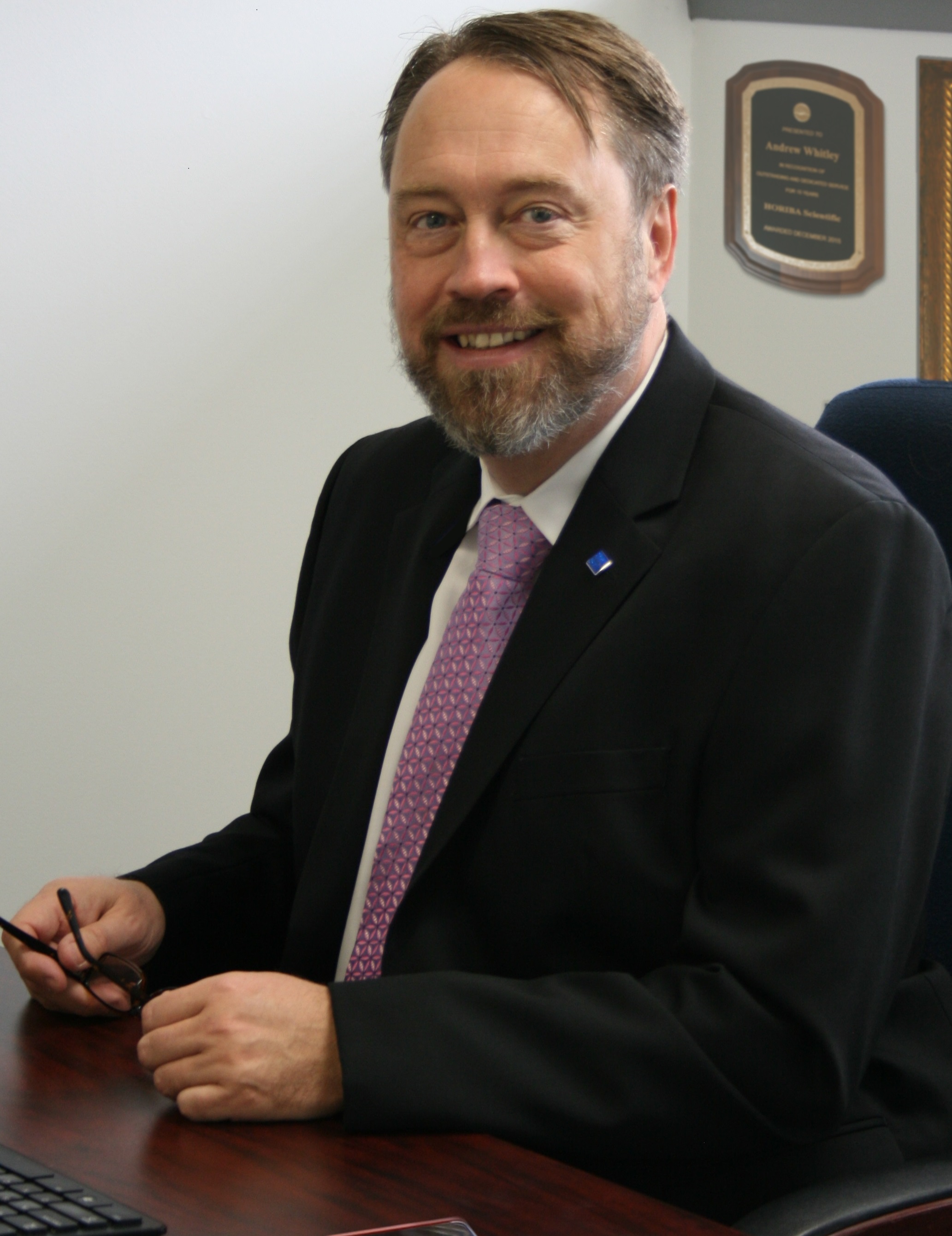
Andrew Whitley, Committee Member
|
Work Title: VP of Business Development
Number of Years as a SAS Member: 26
Biggest benefit of SAS: Networking (partying!)
What did you want to be when you were a child? Striker for Nottingham Forest
What made you fall in love with spectroscopy? Solving real world problems
What is your favorite part of your career or job? Making others succeed
Which scientist, past or present, would you like to meet and why? Alexander Fleming
|
2025 SAS Early Career Travel Grant Recipients
Name: Ailsa Geddis

Affiliation: Université de Montréal
Dr Ailsa Geddis is a Postdoctoral Researcher at the Université de Montréal. She earned her MChem from the University of St Andrews in 2020 and completed her PhD at the University of Edinburgh in 2024. Her research to date has focused on the development of SERS (Surface-Enhanced Raman Scattering) sensor platforms designed to address a broad spectrum of clinical and biological challenges. Building on this foundation, Dr Geddis’s current and future work explores the dynamic intersection of spectroscopy and artificial intelligence. She is particularly interested in applying machine learning to extract subtle, complex patterns from spectral data—using transparent and explainable algorithms to advance the interpretability of biomedical signals.
Beyond the lab, Ailsa is deeply committed to science outreach and public engagement. During her PhD, she was awarded a competitive scholarship to lead and contribute to several outreach initiatives, including the "Workshop in a Box" scheme, which delivered IR and UV spectrometers to remote and underfunded high schools across Scotland to support learning in Forensic and Heritage science. In 2023, she played a leading role in designing and delivering an interactive outreach project on robotic surgery, showcased in the Science Futures tent at Glastonbury Festival.
Dr Geddis is excited about the future of interdisciplinary science and looks forward to the opportunities the coming years will bring.
Title of SciX talk: Exploring Brain Chemistry: Developing SERS-Optophysiology with Optogenetics for Enhanced Neurotransmitter Sensing
Abstract:
In situ detection of neurotransmitters within neuronal networks is crucial for understanding brain function and disease. Chemical gradients around neurons and the extracellular space are critical for maintaining normal biological functions, and their dysregulation can lead to neurological diseases like Parkinson’s disease (PD). Traditional methods such as fluorescence microscopy and electrochemistry have been valuable for studying molecular activity, but they fall short in capturing the dynamic complexity of these systems. Surface-enhanced Raman scattering (SERS) optophysiology with optogenetics is a novel approach pioneered by the Masson and Trudeau groups.1 It makes use of a microneedle functionalised with plasmonic nanomaterials and a laser to provide information on molecular vibrations present in a physiological sample, which is combined with optogenetics to control neuron activity via light-sensitive proteins, enabling both real-time sensing and manipulation of neural processes.
Recent advances in imaging and biomolecule detection are shifting towards the short-wave IR (SWIR) region.2 SERS in biological samples or live tissue when carried out in the SWIR region allows light to penetrate deeper into tissue, reduces background fluorescence and causes less laser damage.3 Our current research is expanding our near IR optimised technology into the SWIR region, aiming to unlock huge potential for in vivo imaging due to this high biocompatibility. Our newly developed SWIR enabled Raman microscope combined with optimisation of the available SERS nanostructures and the incorporation of a novel machine learning (ML) based algorithm1,4 will help to fully realize the potential of this innovative technique. The SWIR-SERS optophysiology will then be carried out on a novel live mouse brain model in development by the Trudeau group. This project is pushing the boundaries of what is possible at the intersection of Raman and chemical gradient sensing in brain research. In the near future, we will test this on Parkinson’s disease models to uncover the complex pathways that drive the neurodegeneration.
References:
1. Lussier, F. et al, ACS Nano 2019, 13 (2), 1403–1411.
2. Zhu, B. et al, Sensors 2024, 24 (11), 3539.
3. Deriu, C. et al, Nanoscale Adv. 2023, 5 (8), 2132-2166.
4. Masson, J.-F. et al, Nat. Nanotechnol. 2023, 18, 111-123.
Name: Willis Jones
Affiliation: Assistant Professor, Department of Chemistry and Biochemistry, University of North Florida
Bio: Will Jones is an assistant professor in the Department of Chemistry and Biochemistry at the University of North Florida. He received his BS from Wake Forest University (2014) and PhD from the University of Florida (2019), both in Chemistry. His work as a graduate student under the mentorship of Dr. Nico Omenetto focused on the improvement and characterization of shot-to-shot noise in laser-induced breakdown spectroscopy (LIBS). He completed a postdoc at Savannah River National Laboratory under the guidance of Dr. Alicia Fessler, focused on practical applications and instrument development using LIBS, Raman spectroscopy, and time-of-flight mass spectrometry. He began his position at UNF (a primarily undergraduate institution located in Jacksonville, FL) in 2022, where he leads a team of eleven UNF undergraduates in his research laboratory. Dr. Jones’ current research interests are centered in the development of novel analytical calibration strategies that broadly benefit trace level analysis by simplifying and speeding up sample preparation and physical measurement processes, improving upon statistical figures of merit, and correcting for sample matrix effects. The methods developed in the Jones lab are developed with versatility in mind, and can be applied to a wide range of analytical instrumentation. Techniques that have recently come out of his laboratory include multi-channel dilution analysis (MCDA), calibration by proxy (CbPx), and multi-wavelength internal standardization (MWIS).
SciX Abstract:
Multi-Wavelength Internal Standardization
Recent developments in trace analyte determinations can largely be described as “multi-signal” methods, in which both axes of calibration curves are measured instrumental signals instead of the traditional relationship between analyte signal and concentration. While multi-signal methods have various benefits, many are limited in sample throughput, have a limited ability to correct for matrix effects, or require each analyte species to have several significant measurable signals. Multi-wavelength internal standardization (MWIS) is a novel strategy that directly addresses the limitations of some of the recently described multi-signal techniques, using multiple emission wavelengths for both analyte species and a suite of internal standards. Each selected analyte wavelength is standardized using all monitored internal standard wavelengths. Proof-of-concept for MWIS was obtained using inductively coupled plasma optical emission spectrometry (ICP-OES). Spike recovery experiments using complex matrices difficult for ICP-OES provided analyte recoveries of approximately 100% with relative standard deviations on the order of 1%. The proposed MWIS method was compared to (and outperformed) traditional calibration strategies, as well as the multi-signal methods multi-energy calibration (MEC) and multi-internal standard calibration (MISC) that it was developed to address. MWIS was validated through the analysis of certified reference materials, with recoveries for all analytes ranging from 90-118%. MWIS offers direct improvement over some of the more recently described multi-signal calibration techniques, resulting in an extraordinarily high number of calibration points even though only two solutions are prepared. The use of multiple internal standard signals allows the strategy to be applied to important analytes that have few suitable emission wavelengths such as As and Pb. In addition, spectral interferences are easily identified and are simple to exclude from the calculation.
2024 SAS Early Career Travel Grant Recipients
Name: Yeran Bai
Affiliation: University of Arizona, Wyant College of Optical Sciences
Dr. Yeran Bai will join the Wyant College of Optical Sciences at the University of Arizona as an Assistant Professor in January 2025. She earned her Ph.D. from the University of Chinese Academy of Sciences and enhanced her expertise with postdoctoral work at Boston University and the University of California, Santa Barbara. During her graduate and postdoc studies, she focused on the development and application of photothermal infrared imaging. Dr. Bai's current and future research interests lie in biomedical optics, particularly in developing advanced vibrational spectroscopic imaging techniques for biomedical applications. She has significantly improved mid-infrared photothermal microscopy imaging in terms of resolution, speed, and specificity, and has applied these advancements to study various biomedical questions, from microbial responses to drugs to metabolic changes in neurological disorders.
Title of SciX talk:
Exploring Metabolism: Advanced Insights from Photothermal Infrared Microscopy in Biomedical Research
Abstract:
Understanding metabolic processes is crucial for unraveling intricate biological systems. Investigating these processes provides key insights into cellular functions and their dysregulation across various pathologies. Optical imaging techniques, recognized for their superior resolution and specificity, have significantly advanced this field. Among these, vibrational spectroscopic imaging, particularly mid-infrared photothermal (MIP) imaging, stands out as a powerful tool. This label-free technique captures inherent chemical bond vibrations and utilizes photothermal effects induced by mid-infrared (IR) absorption with a visible beam, thus enhancing spatial resolution in far-field mid-IR imaging. It enables sub-micrometer and depth-resolved visualization of living cells and entire organisms. In this presentation, I will discuss the recent advancements in MIP technology, with a particular emphasis on its application to metabolic research. I will highlight our studies in areas such as microbiology, stem cell research, and neurodegeneration, demonstrating the transformative potential of MIP technology in paving new paths for groundbreaking discoveries in metabolic research.
Name: Jay Kitt
Affiliation: University of Utah
Jay P. Kitt, is Research Assistant Professor at the University of Utah in the Department of Chemistry. Dr. Kitt received his BS (2012) and PhD (2016) in chemistry from the University of Utah, where he was awarded an NSF-IGERT Research Fellowship, receiving interdisciplinary research training in nanoscience. He recently completed
his MS in Biomedical Informatics, where his research and educational pursuits were supported by an NIH-NLM Postdoctoral Fellowship. His research interests include the use of big data techniques and multivariate analysis for translational research in medical informatics and for analysis of vibrational spectroscopy data in interfacial chemistry.
Dr. Kitt has been recognized internationally for his research by the Coblentz Society with their Graduate Student Award and by the Society for Applied Spectroscopy with the Barbara Stull Graduate Student Award. And for service to the SAS with the Presidential Service Award. At the University of Utah, he was honored with the Cheves T. Walling Award for his dissertation research and with the W.W. Epstein Outstanding Educator Award for his teaching. Dr. Kitt has authored or co-authored 28 peer-reviewed publications, presented or co-authored more than 75 talks and posters at scientific meetings, and presented 14 invited talks and seminars. He is an active participant in the broader scientific community, as a Parliamentarian and Publications Committee member of the Society for Applied Spectroscopy, as a reviewer for 8 scientific journals, as a poster judge at the SciX analytical chemistry conference, and as a regular judge for local school science fairs.
Title of SciX talk:
Self-Modeling Curve Resolution Analysis of Raman Spectra from Mixed Deuterated and Protiated Phospholipid Membranes Reveals Isotopically-Segregated Lipid Domains
Abstract:
Deuterated phospholipids are often employed as “non-perturbing” probes of phospholipid membrane structure in vibrational spectroscopy, allowing resolution of frequency-shifted vibrational modes of deuterated phospholipids in mixed-phospholipid membranes. Calorimetry of deuterated-phospholipid vesicles has shown lower-temperature melting transitions and reduced cooperative melting, in deuterated lipids. A study combining infrared spectroscopy and molecular-dynamics simulations shows evidence that incorporation of deuterated phospholipids into protiated lipid membranes induces phase-segregation, where two distinct melting transitions occur in calorimetric thermograms. In the current work, phase segregation in individual, mixed deuterated and protiated lipid vesicles is investigated by Raman microscopy. Raman spectra of mixed-lipid vesicles, collected as a function of deuterated-lipid fraction, were examined by self-modeling curve resolution, allowing model-free resolution of spectral vectors and corresponding temperature-dependent composition profiles. The resulting spectral vectors point to microdomain formation in the membrane, indicated by Raman modes in CH- and CD-stretching regions. As the CD/CH composition is varied, domain size increases linearly with deuterated lipid fraction. Deuterated and non-deuterated lipid segregation allows short-range vibrational coupling within pure-lipid domains. To study the impact of dipolar coupling on lipid-domain structure, mixed-lipid vesicles were monitored as a function of temperature. Two distinct melting transitions are observed, and the melting-transition-temperature of each lipid is shifted from that of a pure sample. Interestingly, dipolar coupling is observed in each lipid type until completion of the phase transition, suggesting that coupling between neighboring phospholipids may contribute to phase-separation during the transition. These results question the assumption that deuterated phospholipids are non-perturbing and suggest caution using deuterated phospholipids in membrane research.
|