Early Career Travel Grant
The Early Career Travel Grant is awarded to Early Career scientists who demonstrate merit in the field of spectroscopy and/or financial need.
2025 SAS Early Career Travel Grant Recipients

Dr. Ailsa Geddis
Dr. Ailsa Geddis is a Postdoctoral Researcher at the Université de Montréal. She earned her MChem from the University of St Andrews in 2020 and completed her PhD at the University of Edinburgh in 2024. Her research to date has focused on the development of SERS (Surface-Enhanced Raman Scattering) sensor platforms designed to address a broad spectrum of clinical and biological challenges. Building on this foundation, Dr Geddis’s current and future work explores the dynamic intersection of spectroscopy and artificial intelligence. She is particularly interested in applying machine learning to extract subtle, complex patterns from spectral data—using transparent and explainable algorithms to advance the interpretability of biomedical signals.
Beyond the lab, Ailsa is deeply committed to science outreach and public engagement. During her PhD, she was awarded a competitive scholarship to lead and contribute to several outreach initiatives, including the "Workshop in a Box" scheme, which delivered IR and UV spectrometers to remote and underfunded high schools across Scotland to support learning in Forensic and Heritage science. In 2023, she played a leading role in designing and delivering an interactive outreach project on robotic surgery, showcased in the Science Futures tent at Glastonbury Festival.
Dr Geddis is excited about the future of interdisciplinary science and looks forward to the opportunities the coming years will bring.

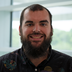
Willis B. Jones
Will Jones is an assistant professor in the Department of Chemistry and Biochemistry at the University of North Florida. He received his BS from Wake Forest University (2014) and PhD from the University of Florida (2019), both in Chemistry. His work as a graduate student under the mentorship of Dr. Nico Omenetto focused on the improvement and characterization of shot-to-shot noise in laser-induced breakdown spectroscopy (LIBS). He completed a postdoc at Savannah River National Laboratory under the guidance of Dr. Alicia Fessler, focused on practical applications and instrument development using LIBS, Raman spectroscopy, and time-of-flight mass spectrometry. He began his position at UNF (a primarily undergraduate institution located in Jacksonville, FL) in 2022, where he leads a team of eleven UNF undergraduates in his research laboratory. Dr. Jones’ current research interests are centered in the development of novel analytical calibration strategies that broadly benefit trace level analysis by simplifying and speeding up sample preparation and physical measurement processes, improving upon statistical figures of merit, and correcting for sample matrix effects. The methods developed in the Jones lab are developed with versatility in mind, and can be applied to a wide range of analytical instrumentation. Techniques that have recently come out of his laboratory include multi-channel dilution analysis (MCDA), calibration by proxy (CbPx), and multi-wavelength internal standardization (MWIS).
2024 SAS Early Career Travel Grant Recipients
The 2024 recipients are Yeran Bai & Jay Kitt
2023 SAS Early Career Travel Grant Recipients
Name: Stephanie Zaleski
Affiliation: Department of Chemistry & Biochemistry, California State University East Bay
Bio: Stephanie Zaleski joined the Department of Chemistry & Biochemistry as an Assistant Professor at CSU East Bay in 2020. She obtained her B.A. in Biochemistry from Barnard College in 2011 and her Ph.D. in Chemistry from Northwestern University in 2016 working in the lab of Richard P. Van Duyne. She is an established researcher in the field of applied spectroscopy for art conservation and museum studies, as well as the application of SERS to chemical sensing problems. To date, Stephanie has published 14 peer-reviewed journal articles, focusing on topics ranging from the study single molecule electrochemistry by SERS to characterizing arsenic sulfide prints by Raman spectroscopy and SEM-EDS to date mid-19th century Japanese woodblock prints more accurately, as well as assessing the degree of deterioration in 19th-century glass by Visible-IR reflectance spectroscopy. Her current research interests at CSU East Bay include studying fundamental analyte-nanoparticle interactions to improve the use of SERS as an applied analytical technique and establishing an art conservation science research working group between universities, museums, and national laboratories in the San Francisco Bay Area.
Talk title: “Developing Surface-Enhanced Raman Spectroscopy Methods for Quantification Of Complex Analyte Mixtures in Plant-Based Extracts”
Abstract: Analytes such as polyphenolic compounds, which are an important metabolic product associated with plant adaptation to stressful conditions. Anthraquinones are an important class of analytes that are found in solutions of natural red dye extracts of plants. Plant-based extracts are complex mixtures; their composition largely depends on plant species, location, or its biochemical expression. SERS is an ideal method to analyte these mixtures due to its inherent chemical sensitivity and specificity. However, despite its relative popularity, SERS is most often used for the detection of 1-2 analytes.
The goal of this research is to develop a more quantitative model of SERS for the identification and quantification of complex mixtures of analytes found in plant-based extracts, such as polyphenolic compounds in pistachio nut and leaf extracts or anthraquinone compounds found in madder root extracts. In addition, this work will allow us to develop a deeper fundamental understanding of analyte-nanoparticle competitive adsorption and surface interactions, which will help improve the use of SERS for quantitative measurements. Our group's approach involves determining the optimal conditions for solution-based SERS using standard solutions representing the main classes of polyphenolics and anthraquinones found in plant extracts. Using the optimized conditions, we have quantified binding affinities of target analytes by fitting concentration-dependent SERS intensities to a linearized Langmuir adsorption isotherm model. We have found that molecular binding affinity is a more important factor in analyte detection than an analyte's inherent Raman scattering cross section. With this data, we have begun to classify molecular analytes with common features that yield various intensities of SERS signals. We are now exploring the application of our results to actual plant-based extracts.
Name: Hunter B. Andrews
Affiliation: Radioisotope Science and Technology Division, Oak Ridge National Laboratory, Oak Ridge, TN 37831
Talk Title: Overview of Laser-Induced Breakdown Spectroscopy Research at Oak Ridge National Laboratory
Abstract: Laser-induced breakdown spectroscopy (LIBS) is an incredibly robust approach to elemental analysis that is capable of probing solids, liquids, gases, and mixed phases such as aerosols. Additionally, this analysis can be done remotely through fiber optics, providing a pathway for elemental monitoring in hazardous environments such as nuclear reactors or radiological hot cells. Oak Ridge National Laboratory (ORNL) is actively developing LIBS systems for monitoring the off-gases from molten salt reactors and is using the same tools to evaluate treatment options for capturing fission gases during reactor operation. For this application, LIBS is tasked with simultaneously monitoring aerosol particles and noble gases. LIBS is also being used to rapidly map advanced nuclear fuel materials, geological samples, and fly ash. In these applications, LIBS provides an avenue to rapidly evaluate elemental composition and correlations, as well as screen materials to identify regions of interest for a more detailed analysis. LIBS is also being used to investigate environmental samples to aid in carbon capture research. In this application, LIBS is used to rapidly evaluate silicon levels in poplar plant samples to be able to identify genotypes that form phytolith-occluded carbon on the leaf surface. This application aims to better understand the relation between silicon levels, plant genotypes, and plant origins in relation to phytoliths to enable bioengineered plants for optimized carbon retention. This presentation will highlight these examples of LIBS applications at ORNL and provide further updates about many of the projects underway.
2022 SAS Early Career Travel Grant
SAS Early Career Interest Group – Travel Awards
Name: Dr. Olga Eremina
Bio: Olga obtained her Ph.D. in Analytical Chemistry from Lomonosov Moscow State University. She is currently a postdoctoral fellow at the University of Southern California in Dr. Cristina Zavaleta’s research lab, where she utilizes SERS-based sensors for monitoring human and environmental health. At SciX 2022, she will be giving the presentation “Breaking Multiplexity Limits of SERS Imaging to Enable Highly Specific Molecular Imaging and Spatial Profiling of Diseased Tissues”. In this talk, she will describe her work with multiplexed Raman imaging to distinguish 26 different SERS nanoparticles with subcellular resolution.
Name: Dr. Malama Chisanga.
Bio: Malama received his Ph.D. in Analytical Chemistry from the University of Manchester. Currently, he is working in Dr. Jean-Francois Masson’s group at the University of Montreal as a postdoctoral fellow. In his research, Malama is using SERS sensors and chemometrics for medical and clinical applications. Malama’s presentation at SciX 2022 is titled “SERS on a chip: Multiplexed detection of cross-reactivity of SARS-CoV-2-specific antibodies in clinical samples”. His talk will focus on multiplexed detection of antibody cross-reactivity using SERS-active tags that are immobilized on a chip within a microfluidic cell.
2021 SAS Early Career Travel Grant
Name: Dr. Julia Gala de Pablo
Affiliation: Department of Chemistry, University of Tokyo
Abstract Title: High-throughput Raman flow cytometry for directed evolution
Abstract: Flow cytometry is an essential tool for single-cell analysis. Fluorescence-flow cytometry allows analyzing thousands of single cells based on their fluorescence signal using fluorescent staining. However, the need for fluorescent labels is problematic due to its low specificity for small biomolecules, cytotoxicity of staining protocols, and autofluorescence interference. Raman spectroscopy obtains a biochemical fingerprint of single cells in a label-free, non-destructive manner. However, its small cross-section results in slow signal acquisition and low throughput, hindering the interrogation of large cell populations. Coherent Raman scattering methods such as coherent anti-Stokes Raman Scattering (CARS) enhance the light-matter interaction, enabling faster acquisitions and allowing high-throughput implementation. We use Fourier-transform CARS (FT-CARS) to obtain a Raman spectrum every 42 µs in the fingerprint region and analyze cells based on their vibrational characteristics. Integration of a rapid-scan FT-CARS spectrometer and a microfluidic device with acoustic focusing enables Raman flow cytometry at 200 cells/s. With this method, we demonstrated high-throughput flow cytometry of various microalgae such as Chromochloris zofingiensis, Euglena gracilis, and Haematococcus lacustris based on their intracellular contents of carbohydrates, proteins, chlorophyll, and carotenoids. We also show that our FT-CARS flow cytometer can characterize differences in metabolic activity among Euglena gracilis clones generated by ion beam mutagenesis. We believe that the combination of ion-beam mutagenesis and Raman flow cytometry opens a new path to metabolic engineering, that is, creating and characterizing cells with specific phenotypes in a label-free manner.
Bio: Dr Julia Gala de Pablo studied a BSc in Physics and a BSc in Biochemistry at the University Complutense of Madrid (Spain). In 2015, she moved to the University of Leeds (UK), defending her PhD in Raman spectroscopy of live single colorectal cancer cells in 2019. She is currently a JSPS postdoctoral fellow at the University of Tokyo in Goda-lab working in Fourier-Transform Coherent anti-Stokes Raman Scattering for flow cytometry and sorting.
Name: Dr. Rupali Mankar
Affiliation: Department of Electrical and Computer Engineering, University of Houston
Abstract Title: Polarization sensitive photothermal mid-infrared spectroscopic imaging of human bone marrow tissue
Abstract: Collagen quantity and integrity play a significant role in understanding diseases such as myelofibrosis (MF). Label-free mid-infrared spectroscopic imaging (MIRSI) has the potential to quantify collagen while minimizing the subjective variance observed with conventional histopathology. Polarization-sensitive Infrared (IR) spectroscopy provides chemical information while also estimating tissue dichroism. Quantitative chemical and structural information can potentially aid MF grading and improve pathological agreement on the diagnosis by quantifying chemical and structural information of collagen fibers. We are presenting the first study of polarization-dependent spectroscopic variations in collagen from human bone marrow samples. We translate polarization-sensitive IR studies in animal models into a clinically viable method for analyzing human clinical biopsies. We developed a new polarization-sensitive optical photothermal mid-infrared (O-PTIR) spectroscopic imaging scheme that enables sample and source independent polarization control. The proposed imaging scheme allows tissue imaging at higher spatial resolution (0.5µm) with reduced imaging time. OPTIR provides 0.5µm spatial resolution, enabling the identification of thin (≈1µm) collagen fibers that were not separable using Fourier Transform Infrared (FTIR) imaging in fingerprint spectral region at diffraction-limited resolution (≈5µm). We also propose quantitative metrics to identify fiber orientation from discrete band images (amide I and amide II) measured under three polarizations. In previous studies, for collagen fiber orientation, mid-IR imaging was collected using IR lights of multiple polarizations in the range of 00 – 1800. Imaging tissue with multiple polarizations is time-consuming, especially at high spatial resolution. Hence, we proposed a clinically viable imaging scheme to quantify collagen fiber orientation by imaging tissue at only two discrete bands and three polarizations (two orthonormal polarization and the third one at 450 from both orthogonal polarizations). The proposed imaging scheme provides sufficient information that can be translated into a quantitative metric using Jone's calculus. However, mid absorbance imaging with two orthogonal polarizations is inadequate for identifying collagen orientation in clinical samples since human bone biopsies contain collagen fibers oriented in multiple directions. Here, we address this challenge and demonstrate that three polarization measurements are necessary and sufficient to resolve orientation ambiguity in clinical bone marrow samples. Our study is also the first to demonstrate the ability to spectroscopically identify thin collagen fibers (≈1µm diameter) and quantify their orientations, critical for accurate grading of human bone marrow fibrosis.
Brief Bio: Rupali Mankar is a postdoctoral fellow at the University of Houston. She holds a Ph.D. from the University of Houston. Her research focuses on combining IR spectroscopy and machine learning to improvise spectroscopy for clinical translation. She was awarded a postdoctoral fellowship award by the National Laboratory of Medicine (NIH-NLM) for her Biomedical Informatics and Data Science field. In her Ph.D. work, she has automated osteosclerosis (one type of bone marrow fibrosis) and currently working on overcoming the diffraction-limited spatial resolution of IR imaging for comprehensive evaluation of bone marrow fibrosis.
|